Anatomy Rib Cage Posterior View - Figure 1 From Introduction To Chest Wall Reconstruction Anatomy And Physiology Of The Chest And Indications For Chest Wall Reconstruction Semantic Scholar / Rib cage anatomy posterior view.
byAdmin•
0
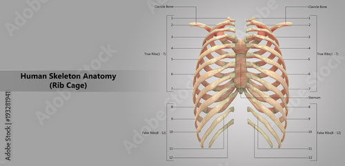
Anatomy Rib Cage Posterior View - Figure 1 From Introduction To Chest Wall Reconstruction Anatomy And Physiology Of The Chest And Indications For Chest Wall Reconstruction Semantic Scholar / Rib cage anatomy posterior view.. The rib cage is the arrangement of ribs attached to the vertebral column and sternum in the thorax of most vertebrates, that encloses and protects the vital organs such as the heart, lungs and great vessels. 16 photos of the rib cage diagram with organs. Diagram of human body, liver rib cage, rib cage diagram labeled, rib cage diagram numbered, rib cage diaphragm, rib cage heart, rib cage organs anatomy, rib cage pain, stomach, diagram of human body, liver rib cage, rib cage diagram labeled, rib cage diagram numbered, rib cage diaphragm, rib cage. Anatomy of the rib cage diagram. Human skeleton system rib cage posterior view anatomy.
It is split into superior and inferior fibres. (b) left lateral chest radiograph (magnified view) obtained at a. Rib cage, in vertebrate anatomy, basketlike skeletal structure that forms the chest, or thorax, and is made up of the ribs and their corresponding attachments to the sternum (breastbone) and the vertebral column. They articulate with the vertebral column posteriorly, and terminate anteriorly as cartilage (known as costal cartilage). The rib below that is rib 2, and it connects to the t2 thoracic vertebra, and so on.
Human Skeleton System Rib Cage With Detailed Labels Anatomy Posterior View Stock Illustration Adobe Stock from as2.ftcdn.net Human skeleton system rib cage anatomy (posterior view) rib cage anatomy of posterior limb and radius view isolated. It is innervated by the first four lumbar nerves, plus the twelfth thoracic nerve. 1278 x 1300 jpeg 105 кб. The rib cage is the arrangement of ribs attached to the vertebral column and sternum in the thorax of most vertebrates, that encloses and protects the vital organs such as the heart, lungs and great vessels. Rib cage anatomy posterior view. Bones of the pelvis and lower back. The articulation with the rib cage leads to regional variations in movement patterns and function (1). The inferior fibres originate from the spinous processes of the t11 to l2 vertebrae and attach to the lower borders of ribs eight to twelve near the angle.
Each are symmetrically paired on a right and left side.
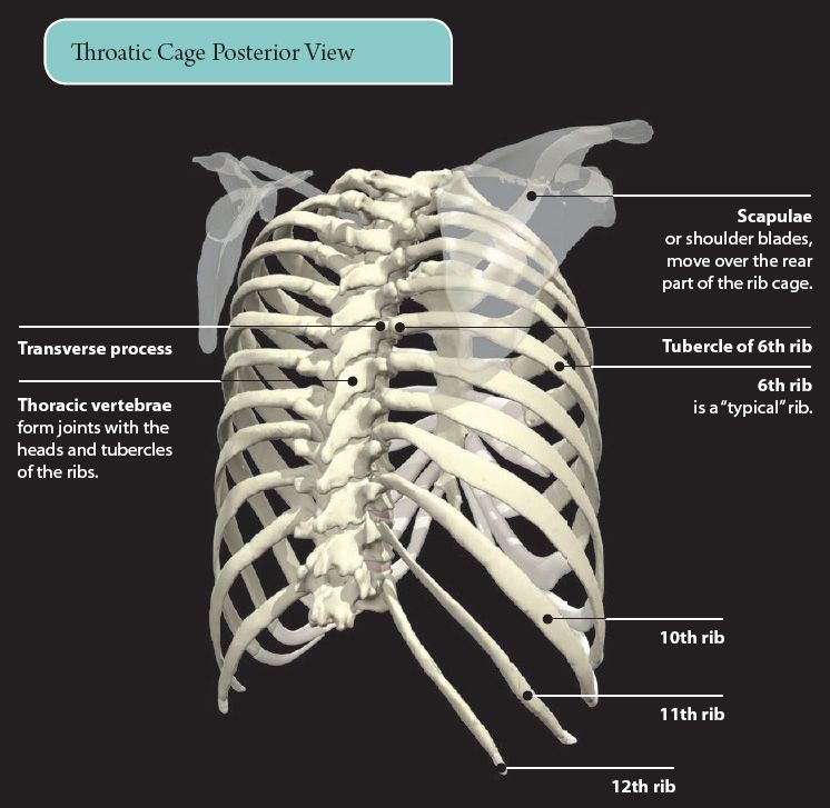
The most superior rib is designated rib 1 and it articulates with the t1 thoracic vertebrae. However, they do not attach directly to the sternum anteriorly, and instead, attach to the. Osteoporosis of the human skeleton, ache of the rib. Our latest youtube film is ready to run. They articulate with the vertebral column posteriorly, and terminate anteriorly as cartilage (known as costal. 1278 x 1300 jpeg 105 кб. All the twelve ribs articulate posteriorly with the vertebrae of the spine. The nomenclature of the costal veins is the same as the arteries. This furrow isn't present in the 11th and 12th ribs. Anatomy of the rib cage diagram. Each pair articulates with a different thoracic vertebra on the posterior side of the body. It may occur after an obvious injury or without explanation. Anatomy of the rib cage diagram.
Scalenus anterior, posterior and medius muscles have attachments on the first and second ribs; At the chest, many rib bones connect to the sternum via costal cartilage,. Unlike a standard chest radiograph, this projection applies a lower kv higher mas technique to highlight bony structures. It may occur after an obvious injury or without explanation. The thoracic spine, composed of 12 segments, is the longest subsection of the vertebral.
4 The Thorax Basicmedical Key from basicmedicalkey.com All ribs are attached posteriorly to the thoracic vertebrae and are numbered accordingly one to twelve. New users enjoy 60% off. (rib cage) anatomy with detailed labels posterior view 3d illustration of human skeleton system (rib cage) anatomy. Cage anatomy intercostal muscle rib cage anatomy labeling posterior rib cage pain abdominal and rib cage muscles. 1278 x 1300 jpeg 105 кб. Extent of the region and the articulations with the rib cage. This furrow isn't present in the 11th and 12th ribs. At the chest, many rib bones connect to the sternum via costal cartilage,.
It depresses the lower rib cage.
The thoracic spine and rib cage yogabody anatomy. Bones of the pelvis and lower back. Extent of the region and the articulations with the rib cage. However, they do not attach directly to the sternum anteriorly, and instead, attach to the. 16 photos of the rib cage diagram with organs. Thus, the posterior ribs are farther from the film and are on the right. Ribs (ap view) the ribs ap view is a specific projection employed in the assessment of the posterior ribs. Download 511 human anatomy skeleton ribcage stock illustrations, vectors & clipart for free or amazingly low rates! It may occur after an obvious injury or without explanation. The vertebral column of the lower back includes the five lumbar vertebrae, the sacrum, and the coccyx. Rib cage anatomy posterior view / image of human skeleton system rib cage bone joints described with labels anatomy posterior view qa433537 picxy. Ribs with veins posterior view. The rib below that is rib 2, and it connects to the t2 thoracic vertebra, and so on.
(b) left lateral chest radiograph (magnified view) obtained at a. Human skeletal system anatomy view. Human muscles · april 17, 2020. Of all 24 ribs, the All ribs are attached posteriorly to the thoracic vertebrae and are numbered accordingly one to twelve.
The Lumbar Vertebrae Are In Human Anatomy The Five Vertebrae Between The Rib Cage And The Pelvis from www.imago-images.de Serratus posterior this muscle is present posteriorly within the thoracic wall. Download 511 human anatomy skeleton ribcage stock illustrations, vectors & clipart for free or amazingly low rates! A rib has a flat body, as you can see from the picture of the anatomy of the human rib cage. Measuring rib cage and abdominal movement is the most common technique for assessing respiratory effort in laboratory. Rib cage anatomy posterior view / image of human skeleton system rib cage bone joints described with labels anatomy posterior view qa433537 picxy. (b) left lateral chest radiograph (magnified view) obtained at a. It depresses the lower rib cage. 1278 x 1300 jpeg 105 кб.
(b) left lateral chest radiograph (magnified view) obtained at a.
The most superior rib is designated rib 1 and it articulates with the t1 thoracic vertebrae. The vertebral column of the lower back includes the five lumbar vertebrae, the sacrum, and the coccyx. The nomenclature of the costal veins is the same as the arteries. It is innervated by the first four lumbar nerves, plus the twelfth thoracic nerve. However, they do not attach directly to the sternum anteriorly, and instead, attach to the. Serratus posterior this muscle is present posteriorly within the thoracic wall. Inserts at the xiphisternum and the 5 th to 7 th costal cartilages; All ribs are attached posteriorly to the thoracic vertebrae and are numbered accordingly one to twelve. Additional variation is associated with ethnicity. (b) left lateral chest radiograph (magnified view) obtained at a. Download 511 human anatomy skeleton ribcage stock illustrations, vectors & clipart for free or amazingly low rates! Anatomy of the rib cage diagram. New users enjoy 60% off.
Inserts at the xiphisternum and the 5 th to 7 th costal cartilages; anatomy rib cage. It often involves two projections, one of the supradiaphragmatic ribs and two of the subdiaphragmatic ribs.